|
Apparently, improvements in MRI technology make it possible to measure lesions without exposing the patient to radiation. Therefore, we believe it's reasonable to ask the medical community if MR imaging can and should be used, especially when frequent monitoring is required. A second safer alternative might be sonograms, in circumstances were approximate monitoring is all that's required.
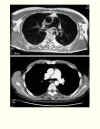 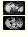 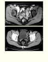 
Click images above to enlarge view of sample
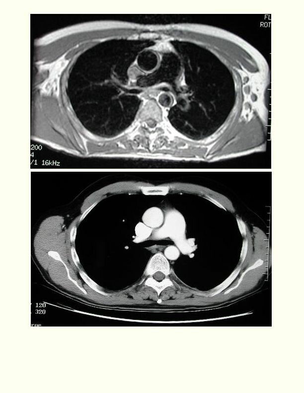
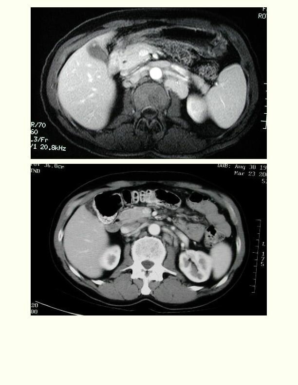
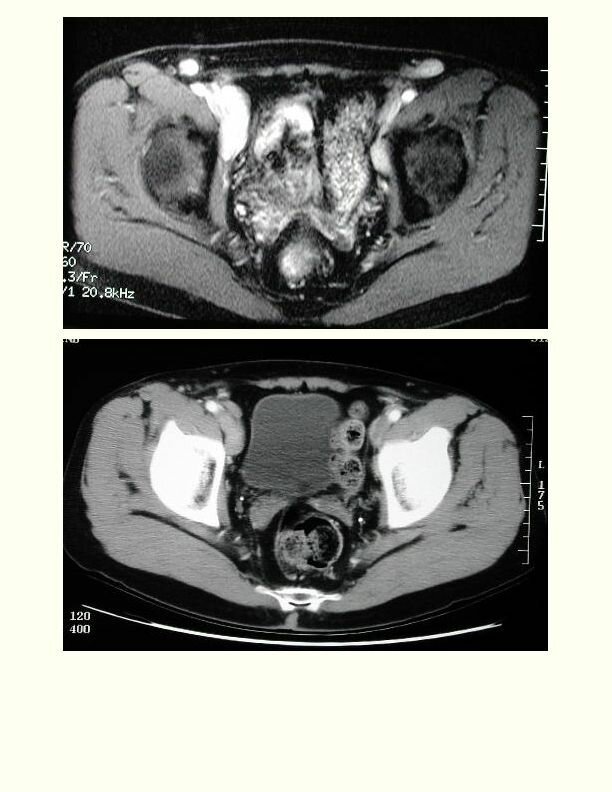
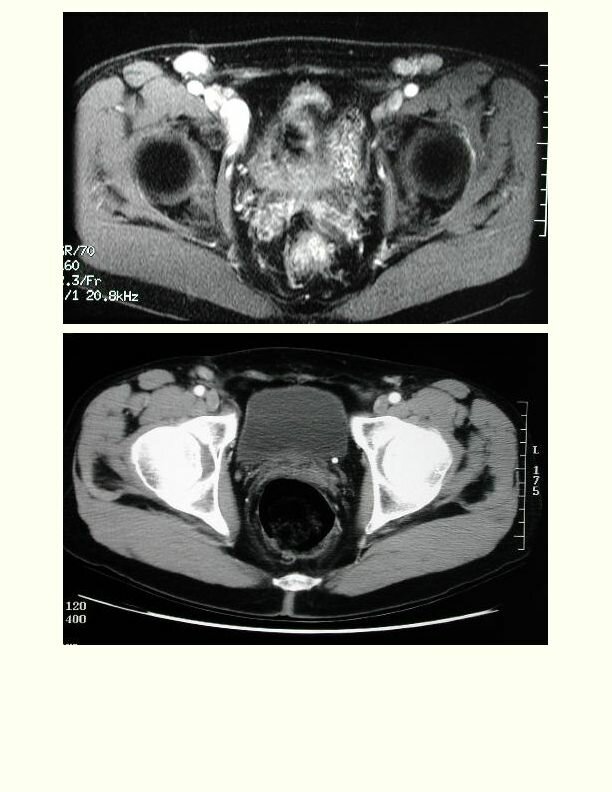
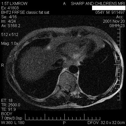
MRI and CT images of same pt. MR images are on top
I am attaching small versions of the CT and MR images. CT 2, 3, 4, 5 correspond with MRI 2,3,4,5.
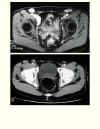
I omitted CT 1 and MR 1 because they did not quite match. I also included 3 nice coronal MR images.
The ability to image in the Coronal plane is an advantage of MRI.
For all the sections, the MRI shows the nodes better than on the CT. - Dr. Russell Low, radiologist
Also see Comparing Images
|